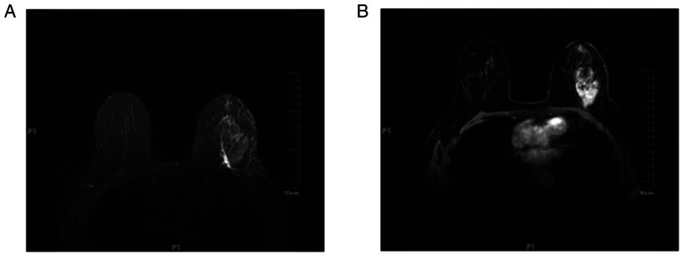
Figure 1.
Magnetic resonance imaging examination of a 62-year-old woman with invasive ductal carcinoma of the left breast with metastasis to the right axillary lymph nodes confirmed by biopsy. (A) The T2-weighted fat suppression sequence revealed the presence of edema around the tumor mass and under the skin. (B) The transverse contrast-enhanced dynamic sequence revealed that there was a markedly irregular enhancement mass in the lateral quadrant at the level of the nipple.

