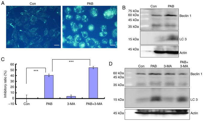
Figure 6.
PAB induces autophagy and inhibits autophagy-mediated cell death. (A) At 24 h, 4 µM PAB increased the positive dots of MDC staining as determined by fluorescence microscopy. Scale bar, 15 µm. (B) The expression levels of Beclin 1 and LC3 following PAB treatment. Representative images of beclin 1, actin and LC3 blots were from different gels, but the same batch of samples. (C) Proliferation inhibition was evaluated by the MTT assay at 24 h following PAB treatment. 3-MA is an autophagy inhibitor. Data are presented as the mean ± SD, n=3. ***P<0.001. (D) The expression levels of beclin 1 and LC3 following PAB or 3-MA treatment at 24 h post-PAB treatment. Representative images of beclin 1, LC3 and actin blots are from different gels, but the same batch of samples. PAB, pseudolaric acid B; MDC, monodansylcadaverine.

