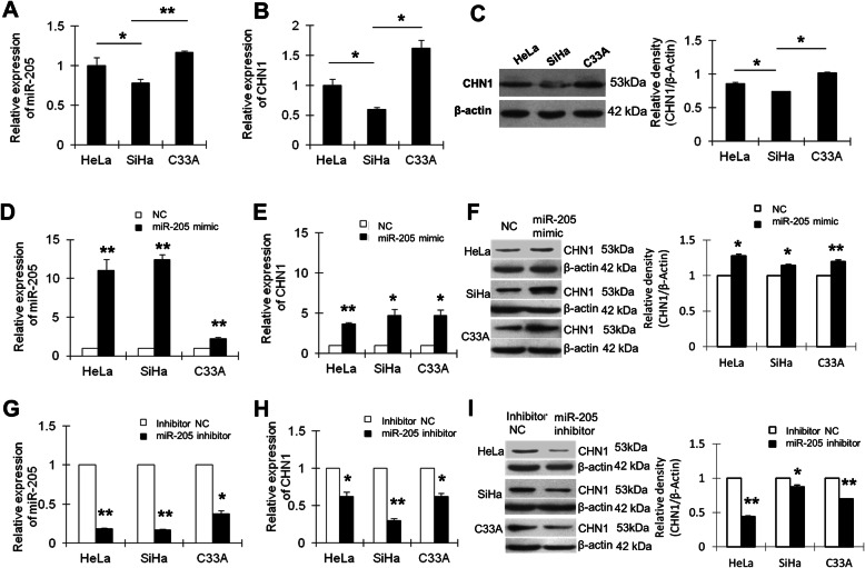
Fig. 3.
Confirmation of the relationship between miR-205 and CHN1. a The expression of miR-205 in HeLa, SiHa, and C33A cells was detected by qRT-PCR. U6 served as an internal reference and was used to normalise miR-205 expression. The y-axis displays the relative expression of miR-205 normalised to the expression of U6. b The expression of CHN1 mRNA in HeLa, SiHa, and C33A cells was detected by qRT-PCR. GAPDH served as an internal reference gene. c CHN1 protein expression was detected by western blotting. β-Actin was used as a loading control. The black histogram shows the optical densities of the signals quantified by densitometric analysis and represented as the CHN1 intensity/β-Actin intensity for normalisation of gel loading and transfer. d HeLa, SiHa, and C33A cells were transfected with NC or miR-205 mimic. The expression of miR-205 was detected by qRT-PCR. e The level of CHN1 mRNA was detected by qRT-PCR. GAPDH served as an internal reference gene. f CHN1 protein expression was detected by western blotting. β-Actin was used as a loading control. g HeLa, SiHa, and C33A cells were transfected with inhibitor NC or miR-205 inhibitor. The expression of miR-205 was detected by qRT-PCR. h CHN1 mRNA expression was detected by qRT-PCR after transfection of cells with inhibitor NC or miR-205 inhibitor. GAPDH served as an internal reference gene. i CHN1 protein expression was detected by western blotting after transfection of cells with inhibitor NC or miR-205 inhibitor. β-Actin was used as a loading control. *P < 0.05, **P < 0.01. The cropping of the blot was done. Full-length uncropped blots are presented in Supplementary Figure 1, which all the samples derived from the same experiment and blots were processed in parallel

