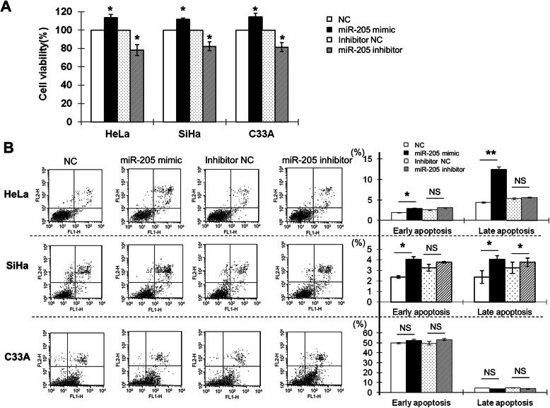
Fig. 4.
The effects of miR-205 on the proliferation and apoptosis of human cervical cancer cells. a HeLa, SiHa, and C33A cells were transfected with the NC, miR-205 mimic, inhibitor NC, or miR-205 inhibitor. At 48 h after transfection, cell proliferation was determined by CCK-8 assay. All experiments were performed at least three times, and cell proliferation was determined as the stimulation index (SI; i.e., the ratio of absorbance at 450 nm of cells transfected with miR-205 mimic or inhibitor to that of cells transfected with NC or inhibitor NC). b HeLa, SiHa, and C33A cells were transfected with the NC, miR-205 mimic, inhibitor NC, or miR-205 inhibitor for 48 h. Cells were then stained with annexin V/PI and subjected to flow cytometry analysis. Lower left quadrant, viable cells (annexin V-FITC and PI negative); lower right quadrant, early apoptotic cells (annexin V-FITC positive and PI negative); upper right quadrant, late apoptotic/necrotic cells (annexin V-FITC and PI positive). The average percentage of apoptotic cells was analysed in cells transfected with miR-205 mimic or inhibitor at early and late stages. The histograms represent the average percentages of apoptotic cells in cells transfected with miR-205 mimic at early and late stages or miR-205 inhibitor at early and late stages. The experiment was repeated at least three times. *P < 0.05, **P < 0.01, NS: not significant

