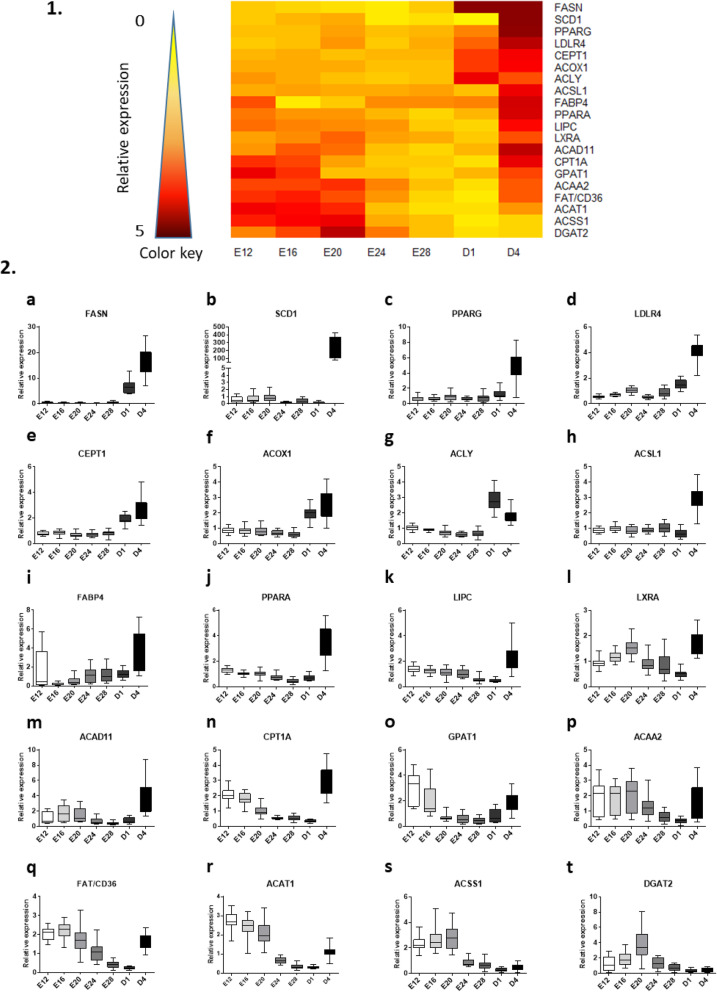
Fig. 3.
Relative hepatic expression of lipid-related genes from E12 to D4. 1. Heatmap illustration of liver gene expressions at different stages in mule ducks. Low gene expression is indicated in yellow, while high expression is in red, according to the color key. 2. Box-and-whisker plots representations of expression profile of FASN (a), SCD1 (b), PPARG (c), LDLR4 (d), CEPT1 (e), ACOX1 (f), ACLY (g), ACSL1 (h), FABP4 (j), PPARA (j), LIPC (k), LXRA (l), ACAD11 (m), CPT1A (n), GPAT1 (o), ACAA2 (p), FAT/CD36 (q), ACAT1 (r), ACSS1 (s), DGAT2 (t) in the liver of mule duck during development. The boxes extend from the 25th to the 75th percentiles, and the whiskers range from the lowest value to the highest

