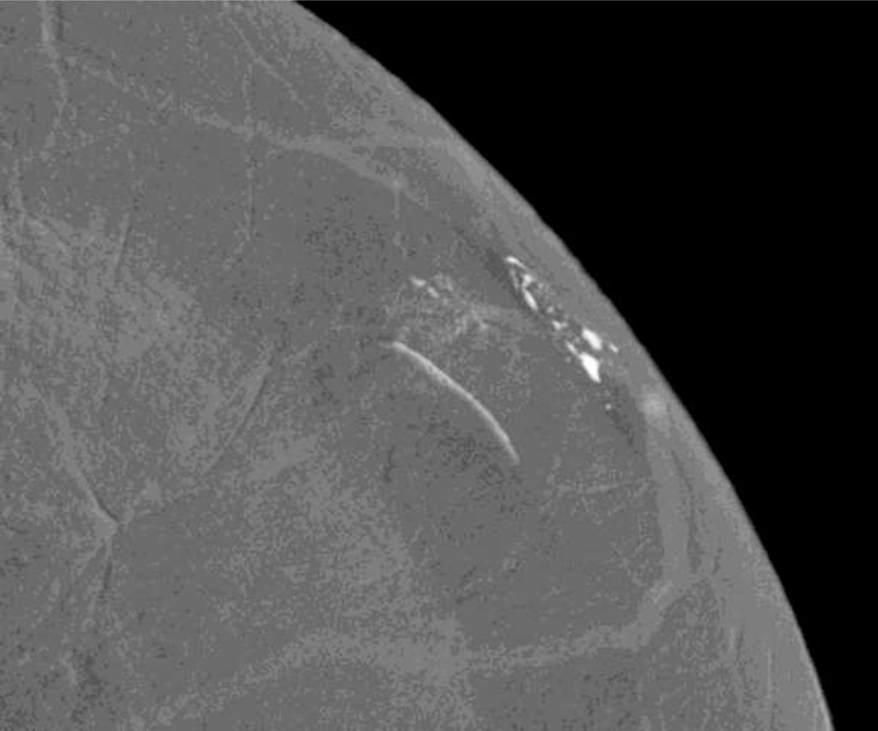
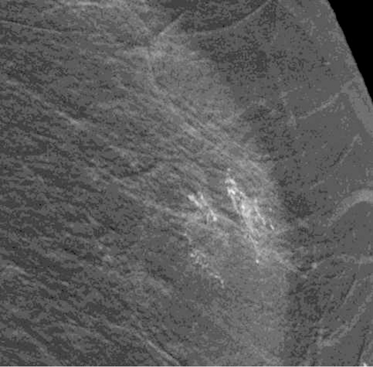
Fig. 5—
Left mammogram of Patient E, a 59-year-old female with right breast abnormality on screening mammogram, which was subsequently biopsied yielding Invasive Ductal Carcinoma and DCIS. Recombined images of the left breast CEDM demonstrate apparent linear non-mass enhancement in a superficial location in the outer (A) upper (B) quadrant.


