Figure 2.
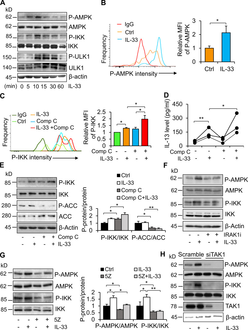
AMPK is activated by IL-33, and feedback inhibits IL-33 signaling in ILC2. (A) Phosphorylation of AMPK at Thr172 and ULK1 at Ser317, as well as phosphorylation of IKKα/β at Ser176/180, was stimulated by treatment with 10 ng/ml IL-33 in a time-dependent manner in primary differentiated Th2 cells. Cells were cultured in a fresh RPMI medium with 1% FBS for 1 h and then treated with 10 ng/ml IL-33 for 5, 10, 15, 30, and 60 min. Two mice were used for each independent experiment. Three times independently. (B) IL-33 treatment stimulated phosphorylation of AMPK at Thr172 in primary adipose-resident ILC2s. Three to four mice were used for each independent experiment. Data are presented as means ± SEM. *, P < 0.05, Student’s t test. Each experiment was conducted three times independently. (C) Treatment with 1 µM compound C enhanced IL-33–stimulated phosphorylation of IKKα/β in primary ILC2s. Flow cytometry was used for the analysis of ILC2 in B and C. Four to six mice were used for each independent experiment. Data are presented as means ± SEM. *, P < 0.05, Student’s t test. Each experiment was conducted three times independently. (D) Treatment of compound C elevated IL-33–induced secretion of IL-13 in primary ILC2s isolated for eWAT. Four to six mice were used for each independent experiment. *, P < 0.05; **, P < 0.01; Student’s t test. Each experiment was conducted three times independently. (E) Inhibiting AMPK by treatment of 1 µM compound C increased IL-33–stimulated IKKα/β phosphorylation in Th2 cells. ACC, acetyl-CoA carboxylase; p-ACC, phosphorylation of acetyl-CoA carboxylase. One mouse was used for each independent experiment. Data are presented as means ± SEM. *, P < 0.05; **, P < 0.01; Student’s t test. Each experiment was conducted three times independently. (F and G) Inhibiting IRAK1 (F) or inhibiting TAK1 (G) significantly suppressed IL-33–stimulated phosphorylation of AMPK in Th2 cells. Starved cells were treated with 10 µM IRAK1/4 inhibitor or 10 nM TAK1 inhibitor 5Z for 1 h, followed by treatment with 10 ng/ml IL-33 for 10 min. One mouse was used for each independent experiment. Data are presented as means ± SEM. *, P < 0.05; **, P < 0.01. Student’s t test. Each experiment was conducted three times independently. (H) Suppressing TAK1 using siRNA alleviated IL-33–stimulated phosphorylation of AMPK and IKKα/β in Th2 cells. Starved cells were treated with 10 ng/ml IL-33 for 10 min. One mouse was used for each independent experiment. Each experiment was conducted three times independently. Ctrl, control; MFI, mean fluorescence intensity; P, phosphorylation.

