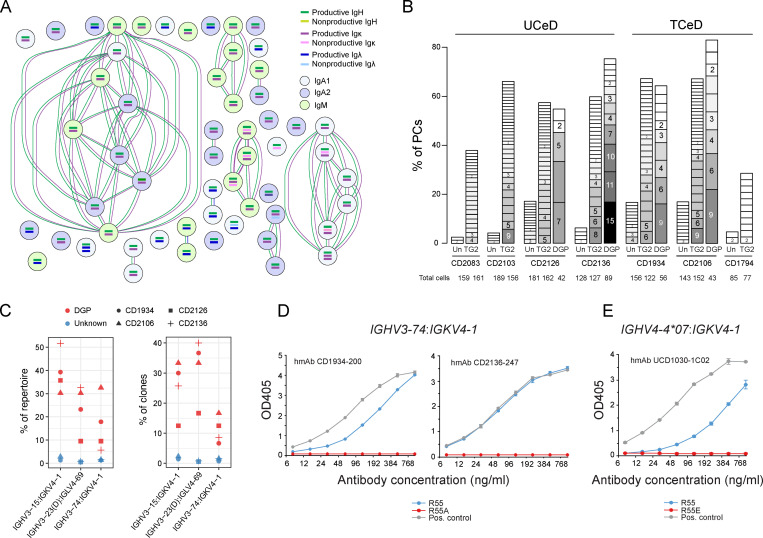
Figure 4.
Clonal expansion, repertoire analysis, and site-directed mutagenesis of antibodies of PCs in CeD. (A) Example of clonal expansion of DGP-specific PCs from a TCeD patient (CD1934) as inferred by BraCeR. Circles represent individual cells, and connections indicate clonal relationship based on both heavy and light chains. (B) Clone group sizes for each specificity in each CeD patient shown as percentage of PCs. Darker shades of gray and larger boxes indicate bigger clones, with number of cells in each clone group shown. Clones with two or more cells are shown. Un, unknown specificity. (C) Percentage of total DGP-specific PCs (left) or DGP-specific clone groups (right) in each patient using the three dominating V-gene chain pairings. IGHV3-23 and IGHV3-23D are collectively referred to as IGHV3-23(D). (D and E) Importance of heavy chain R55 for DGP binding assessed by ELISA. Two patient-derived DGP-specific hmAbs using IGHV3-74:IGKV4-1 reported in this study (D) and one previously reported DGP-specific hmAb (UCD1030-1C02; Steinsbø et al., 2014) using IGHV4-4*07:IGKV4-1 (E) were analyzed with or without R in position 55. A previously characterized DGP-specific hmAb (UCD1002-1E03; Steinsbø et al., 2014) using IGHV3-15:IGKV4-1 was used as positive control for comparison. Error bars indicate SD based on sample duplicates.

