
Figure 2.
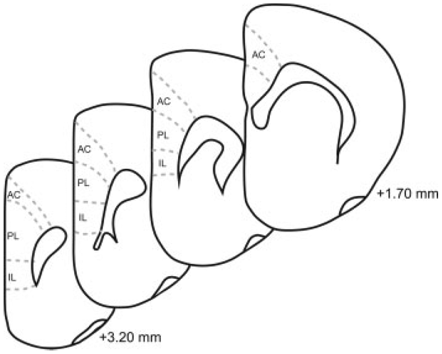
Schematic diagram of coronal sections through prefrontal cortex. The cortical regions (anterior cingulate, AC; prelimbic; PL; infralimbic, IL) from which samples were taken are shown. Coordinates indicate position relative to bregma (Paxinos and Watson, 1998).