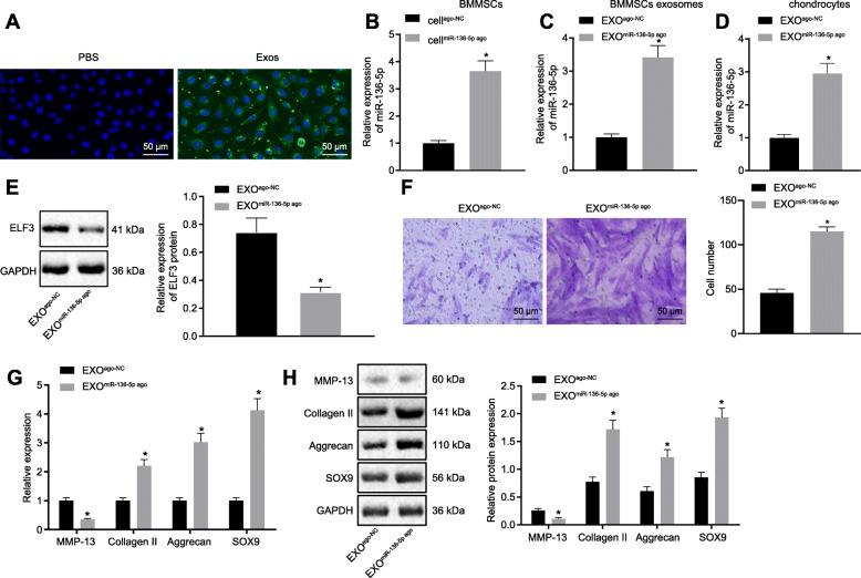
Fig. 4.
The migration and ECM secretion of chondrocytes were promoted by miR-136-5p-exo. a A typical immunofluorescence pattern of DIO (green)-labeled exosomes absorbed by chondrocytes, whose nuclei were stained with DAPI (blue) (× 200). b The expression of miR-136-5p in transfected BMMSCs examined by RT-qPCR. c The expression of miR-136-5p in exosomes secreted from transfected BMMSCs examined by RT-qPCR. d The expression of miR-136-5p in chondrocytes after exosome treatment examined by RT-qPCR. e Western blot analysis of the expression of ELF3 in chondrocytes treated with exosomes. f Migration analysis of chondrocytes (× 200). g The mRNA expressions of MMP-13, collagen II, aggrecan, and SOX9 in chondrocytes examined by RT-qPCR. h Western blot analysis of the protein expressions of MMP-13, collagen II, aggrecan, and SOX9 in chondrocytes. Asterisk symbol indicated comparison with the EXOago-NC or cellago-NC group. A value of p < 0.05 was considered to be statistically significant. The above data were measurement data and expressed as mean ± standard deviation. The independent sample t test was used for comparison between the two groups. One-way ANOVA was used among multiple groups, followed by Tukey’s post hoc test

