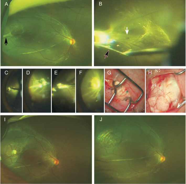
Figure 1. Case 1, a 7-year-old boy diagnosed as PHPV with temporal RD at the right eye.
A: Preoperative fundus photo. Black arrow shows the vitreous traction. B: Small retinal hole was found beneath the vitreous traction under the microscopy. C-H: Key steps of the surgical procedures; C: Coagulation of the vitreous traction; D: Cutting the vitreous traction; E: Trimming the traction by vitrectomy cutter; F: Cryocoagulation under microscopy; G, H: Minimal invasive conjunctival incision and silicone scleral buckle. I, J: Retinal reattachment with temporal ridge 2-day and 1-month after surgery, respectively.

