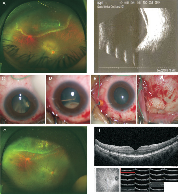
Figure 3. Case 4, a 45-year-old woman with superior RD at the right eye and high myopia of both eyes and had a history of retinal photocoagulation at the right eye.
A: Preoperative fundus photo showed RD with retinal holes and vitreous traction. Laser spot was seen around the posterior margin of the RD area. B: B-scans of the right eye preoperatively. C-F: The key steps of the surgical procedures. C, D: Trimming the vitreous traction around the retinal holes by vitrectomy cutter; E: Injecting the viscoelastic solution into the vitreous cavity to maintain the ocular pressure; F: Minimal invasive conjunctival incision and silicone scleral buckle. G: Postoperative fundus photo showed retinal reattachment. H: Postoperative OCT showed normal macula.

