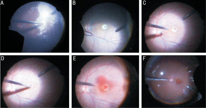
Figure 1. The patient who suffered with rhegmatogenous retinal detachment primary with macular hole (diameter=341 µm, right eye) had underwent previous vitreoretinal surgeries twice. The first surgery was vitrectomy and ILM peeling with silicone oil for tamponade, and then the hole closed. Following silicone oil extraction combined with phacoemulsification and IOL implantation was performed 6mo later. However, recurrent macular hole with diameter of 818 µm was discovered at 1wk after the second surgery.
A: Triamcinolone acetonide was used for staining and identifying whether there was residual vitreous gel; B: Little left ILM could be seen by indocyanine green staining; C: The flute needle was put onto the hole edge to make a cuff of MH with passive suction and slightly blow; D: Gently massage on the parafoveal retina toward the center with vit probe; E: Autologous whole blood was injected into vitreous cavity and the macular hole was fully covered; F: Thorough fluid-air exchange was performed to drain off the liquid of vitreous cavity.

