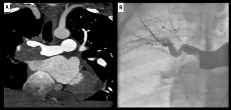
Figure 1. (A) CTPA is showing large flling defect in the distal right pulmonary artery causing subtotal occlusion of the right upper lobar branch and total occlusion of the middle and lower right lobar branches with free fow of contrast through the main and lef pulmonary arteries.
(B) Selective pulmonary angiography is showing flling defect in the distal right pulmonary artery with subtotal occlusion of the right upper lobar branch and amputation of middle and lower right lobar branches. CTPA, computed tomography pulmonary angiography.

