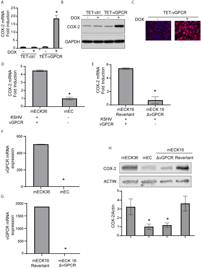
Fig 3. Use of full KSHV genome bearing cells to analyze COX-2 expression regulation by vGPCR.
A) Fold-changes of COX-2 mRNA expression determined by RT-qPCR in Tetracycline-inducible vGPCR (TET-vGPCR) and control mECK36 cells stimulated with doxycycline for 24 hours. COX-2 mRNA was measured in triplicate and is presented as means ± SD. (*P<0.05). B) COX-2 protein expression levels were determined by immunoblotting in Tetracycline-inducible vGPCR (TET-vGPCR) and control mECK36 cells stimulated with doxycycline for 24 hours. GAPDH was used as a loading control. C) IFA for COX-2 (red) in Tetracycline-inducible vGPCR (TET-vGPCR) and control mECK36 cells stimulated with doxycycline for 24 hours. Cell nuclei were counterstained with DAPI (blue). D) Fold-changes of COX-2 mRNA expression determined by RT-qPCR in mECK36 and mEC cells (originated from the former and generated by selection of those that have lost the KSHVBAC36). COX-2 mRNA was measured in triplicate and is presented as means ± SD. (*P<0.05). E) Fold-changes of COX-2 mRNA expression determined by RT-qPCR in mECK16 derived Δ-vGPCR or revertant virus (see Materials and methods). COX-2 mRNA was measured in triplicate and is presented as means ± SD. (*P<0.05). F) vGPCR mRNA expression determined by RT-qPCR in mECK36 and mEC cells (KSHV-negative cells originated from the former and generated by selection of those that have lost the KSHVBAC36). The lowest CT value obtained in KSHV-negative mEC cell samples was assigned as the limit of detection for vGCPR expression. vGPCR mRNA was measured in triplicate and is presented as means ± SD. (*P<0.05). G) vGPCR mRNA expression determined by RT-qPCR in mECK16 derived Δ-vGPCR or revertant virus (see Materials and methods). The lowest CT value obtained in mECK16 Δ-vGPCR cell samples was assigned as the limit of detection for vGCPR expression. vGPCR mRNA was measured in triplicate and is presented as means ± SD. (*P<0.05). H) COX-2 protein expression levels were determined by immunoblotting in mECK36, mEC and mECK16 derived Δ-vGPCR or revertant virus (see Materials and methods). COX-2 protein levels were measured in triplicate and are presented as means ± SD. (*P<0.05).

