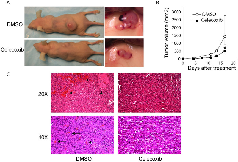
Fig 6. vGPCR-transformed cells tumorigenicity is inhibited by Celecoxib treatment.
A) Mice (n = 7) were injected S.C. with vGPCR-NIH3T3 cells and treated with the COX-2 inhibitor Celecoxib I.P. or vehicle (DMSO) three times/week. On the left side, images of mice treated (lower) or not (upper) with Celecoxib at day 15 of treatment. Tumor from non-treated (upper) or treated with Celecoxib (lower) mice are shown in detail on the right. B) The plot shows the growth of tumor volume during the time of treatment (mean +/-range). Higher deviation in the last day for untreated animals reflect the presence of animals with very large tumors by that day observed in all the experiments. Mice were treated with Celecoxib (black circles) or vehicle (white circles). Tumor size was significantly lower in Celecoxib treated samples at all points of the time course (*P<0.05). C) Histological examination of the tumors. Sections of tumors coming from mice, treated with Celecoxib or not (DMSO) as a control, were stained with Hematoxylin-Eosin. Pictures were taken at 20x or 40x magnification. Black arrows in left panels indicates large areas of hemorrhage and necrosis.

