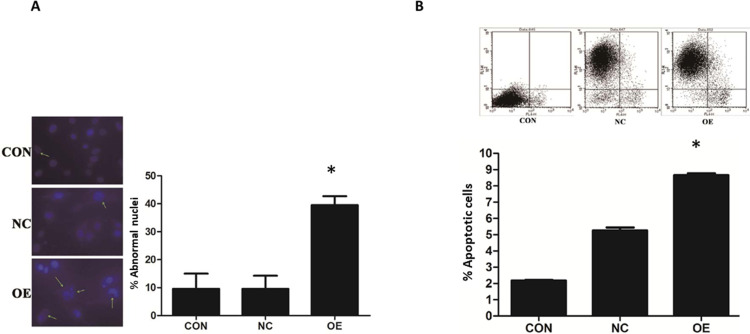
Figure 3.
MiR-16 induction of apoptosis in U87 cells. (A) DAPI staining was utilized to observe the heterotypic cell nucleus and morphological changes of apoptosis. The atypical nuclei are arrowed. The percentage of abnormal nuclei in each group was quantified and compared. *P<0.05 comparing to CON and NC groups. (B) Annexin V-APC staining was used to validate the induction of apoptosis in U87 by miR-16. The positively stained cells were quantified using flow cytometry to calculate the percentage of apoptotic cells. *P<0.05 compared to CON and NC groups. Shown are the representative data sets of three independent experiments.

