Figure 2.

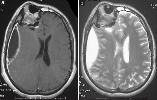
Axial brain magnetic resonance imaging weighted T1 with gadolinium (a) and weighted T2 (b), demonstrating a right frontoparietal compressive subdural empyema associated to giant right fronto-ethmoidal osteoma

Axial brain magnetic resonance imaging weighted T1 with gadolinium (a) and weighted T2 (b), demonstrating a right frontoparietal compressive subdural empyema associated to giant right fronto-ethmoidal osteoma