. 2020 Oct 13;9:e59469. doi: 10.7554/eLife.59469

Figure 2. The Reissner fiber is required for the spontaneous calcium variations of ventral CSF-contacting neurons.

(A) 28–30 hpf embryos expressing the GCaMP5G calcium reporter in CSF-contacting neurons were imaged on the lateral side. Tg(pkd2l1:GCaMP5G) embryos label both dorso-lateral (dl, above the dotted line) and ventral (v, below the dotted line) CSF-contacting neurons (arrowheads). Scale bar: 30 µm. (B) Representative traces of calcium variations in individual ventral CSF-contacting neurons in wild-type (scospondin+/+), heterozygous (scospondinicm15/+) and scospondinicm15/icm15 mutants. Sample traces from individual cells with integral ΔF/F values ranging around the median distribution of the imaged population are represented for each genotype (n = 11 cells). (C) Quantification of the normalized integrated calcium variation over time of ventral CSF-contacting neurons in wild-type (+/+), heterozygous (icm15/+) and scospondinicm15/icm15 mutants (icm15/icm15, blue). Data were collected from five independent experiments and include 10 wild-type embryos (n = 146 cells), 20 heterozygous embryos (n = 287 cells) and 21 scospondinicm15/icm15 mutants (n = 307 cells). Each point represents a single cell. Bottom and top edges of the boxes indicate the 1st and 3rd quartiles. Bold lines represent the median value for each distribution. ns p>0.05, ***p<0.001 (Kolmogorov-Smirnov test). (D) Immunohistochemistry for Pkd2l1 (magenta) and GFP (blue) in Tg(pkd2l1:GCaMP5G) embryos at 30 hpf in the spinal cord of a control sibling (left) and scospondinicm15/icm15 mutant (right). Scale bar: 30 µm. Magnification of the area delineated by dotted line boxes is represented for each condition (r: rostral, c: caudal, d: dorsal, v: ventral). Scale bar: 10 µm. scospondinicm15/icm15 embryos show a similar localization of the Pkd2l1 protein at the developing apical extension (arrowheads) of the CSF-cNs (labeled by the GFP antibody, blue) compared to control siblings. (E) In vivo voltage-clamp recordings from CSF-contacting neurons in the Tg(pkd2l1:GAL4;UAS:mCherry) line at 30 hpf in control embryos (left) and scospondinicm15/icm15 mutants (right). Note the extensive number of events in both conditions (top traces). Bottom traces represent higher temporal magnifications and allow distinguishing single channel openings. See also Figure 2—figure supplement 1 and Figure 2—video 1.

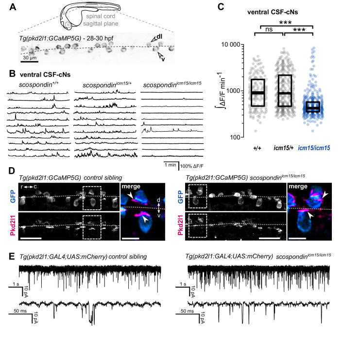
Figure 2—source data 1. Data for Figure 2C and Figure 2—figure supplement 1B–C.

Figure 2.

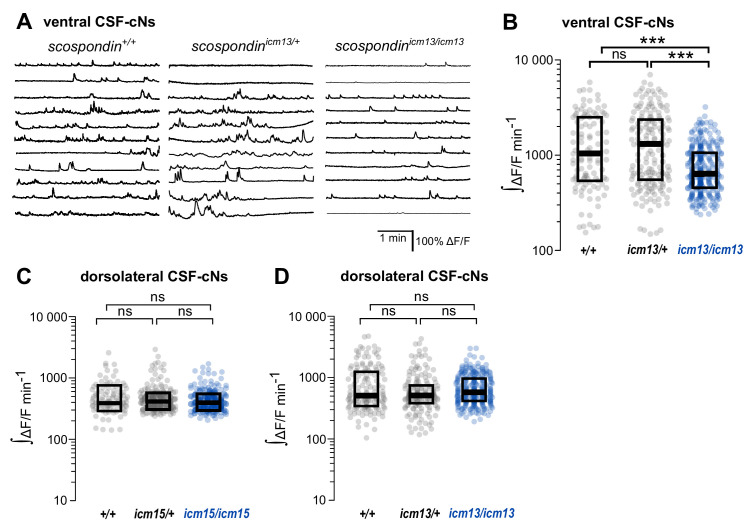
Figure 2—figure supplement 1. The spontaneous activity of dorso-lateral CSF-contacting neurons does not require the Reissner fiber in the embryo.

Figure 2—figure supplement 1.

(A) Traces of calcium variations in ventral CSF-contacting neurons at 30 hpf in wild-type (scospondin+/+), heterozygous (scospondinicm13/+) and scospondinicm13/icm13 null mutants. Sample traces from individual cells with integral ΔF/F values ranging around the median distribution of the imaged population are represented for each genotype (n = 11). (B) Quantification of the normalized integrated calcium variation over time of ventral CSF-contacting neurons in wild-type (+/+), heterozygous (icm13/+) and scospondinicm13/icm13 mutants (icm13/icm13, blue). Data were collected from three independent experiments and include eight wild-type embryos (n = 107 cells), 13 heterozygous embryos (n = 176 cells) and 22 scospondinicm15/icm15 mutants (n = 277 cells). Each point represents a single cell. Bottom and top edges of the boxes indicate the 1st and 3rd quartiles. Bold lines represent the median value for each distribution. ns p>0.05, ***p<0.001 (Kolmogorov–Smirnov test). (C, D) Quantification of the normalized integrated calcium variations over time of dorsolateral CSF-contacting neurons in 30 hpf embryos obtained from scospondinicm15/+ incrosses (C) and scospondinicm13/+ incrosses (D) n = 95, 243, and 255 cells in wild-type, heterozygous and scospondinicm15/icm15 embryos, respectively (C); n = 141, 176, and 168 cells in wild-type, heterozygous, and scospondinicm13/icm13 embryos, respectively (D). ns p>0.05 (Kolmogorov-Smirnov test).
Figure 2—video 1. Intracellular calcium transients of ventral CSF-contacting neurons are reduced in scospondin mutants.
Download video file (1.1MB, mp4)
Sagittal views of the spinal cord of Tg(pkd2l1:GCaMP5G) embryos at 30 hpf. scospondin+/+ (top), scospondinicm15/+ (middle) and scospondinicm15/icm15 siblings (bottom) are represented. In wild-type and heterozygous embryos, ventral CSF-contacting neurons are more active that dorso-lateral ones. Note the sharp decrease in calcium variations of ventral CSF contacting neurons in the homozygous mutant embryo compared to wild-type and heterozygous counterparts. Data were collected at 4 Hz and displayed at 80 Hz. Scale bar: 30 µm.