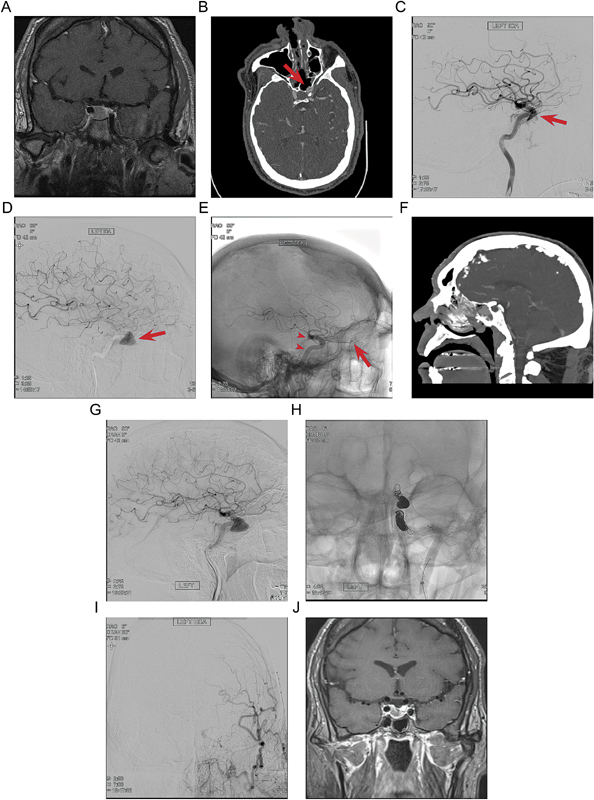
Fig. 1.

( A ) Preoperative contrast-enhanced MRI in the coronal plane demonstrates an enhancing sellar mass. ( B ) Postoperative axial CT angiography demonstrates a bulge of the left ICA in the sphenoid sinus through a bony defect (red arrow). ( C ) Postoperative lateral angiogram of the left ICA shows opacification of the cavernous sinus in the mid-arterial phase, indicating a direct cavernous carotid fistula (red arrow). ( D ) Three-day postoperative lateral left ICA angiogram shows growth of the pseudoaneurysm (red arrow) and diminution of the direct cavernous carotid fistula. ( E ) Magnified unsubtracted lateral angiogram on postoperative day 3 shows the placement of two flow diversion embolization devices across the pseudoaneurysm (red arrowheads), with persistent flow through the ophthalmic artery (red arrow ) . ( F ) Sagittal CT angiogram on postoperative day 5 shows continued filling of the large pseudoaneurysm in the sphenoid sinus. ( G ) Lateral angiogram on postoperative day 6 shows persistent filling of the large pseudoaneurysm in the sphenoid sinus. ( H ) Unsubtracted AP angiogram shows successful platinum coil embolization of the ICA pseudoaneurysm. ( I ) ECA injection angiogram in AP projection on postoperative day 22, taken following after an episode of epistaxis, demonstrates prominent ECA flow via ophthalmic collaterals without filling of the pseudoaneurysm. ( J ) Two-month follow-up contrast-enhanced MRI in the coronal plane demonstrates reduction in the size of the pituitary adenoma. CT, computed tomography; ECA, external carotid artery; ICA, internal carotid artery; MRI, magnetic resonance imaging.
