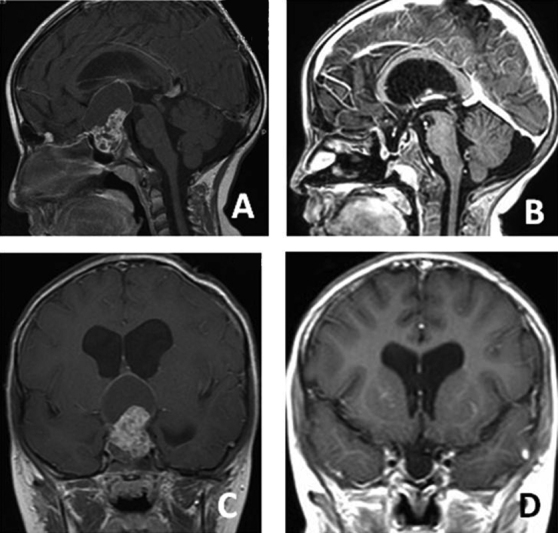
Fig. 5.

A 5-year-old boy presented with visual problems. Preoperative axial ( A ), preoperative sagittal ( B ), postoperative axial ( C ), and postoperative sagittal ( D ) magnetic resonance imaging sections. The pathology was craniopharyngioma. A semipneumatized sphenoid sinus was observed in this patient. Drilling was then required.
