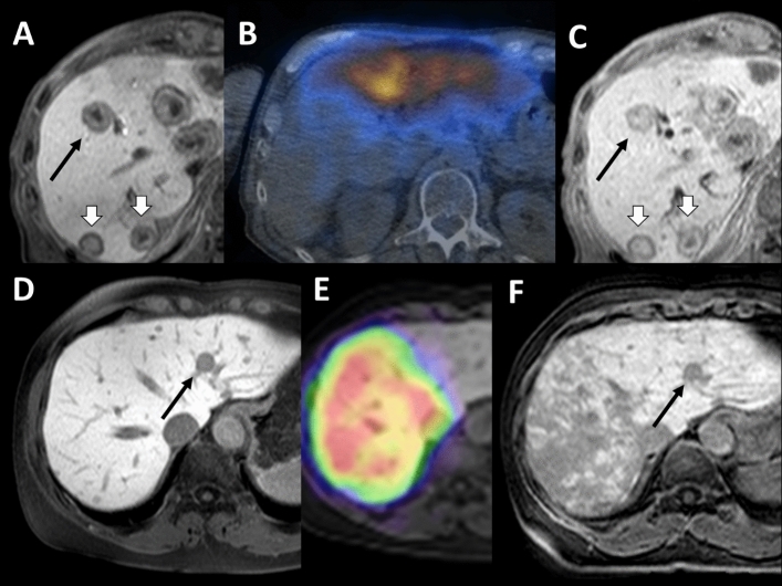
Fig. 4.
Two cases of size regression < 30%, but noticeably smaller, A–C nasopharyngeal carcinoma, D–F breast cancer: A and C) black arrow show a metastasis decreasing from 22 to 20 mm (i.e., 10%), white arrows indicate progressing lesions, e.g., 18–20 mm and 19–22 mm. B Bremsstrahlung-SPECT/CT examination: RE left and segment 4b. Untreated segments 5–8 and 4a. D and E black arrow: 12–10 mm (~ 20%). F Radiation damage in the liver, typical RE image for breast cancer

