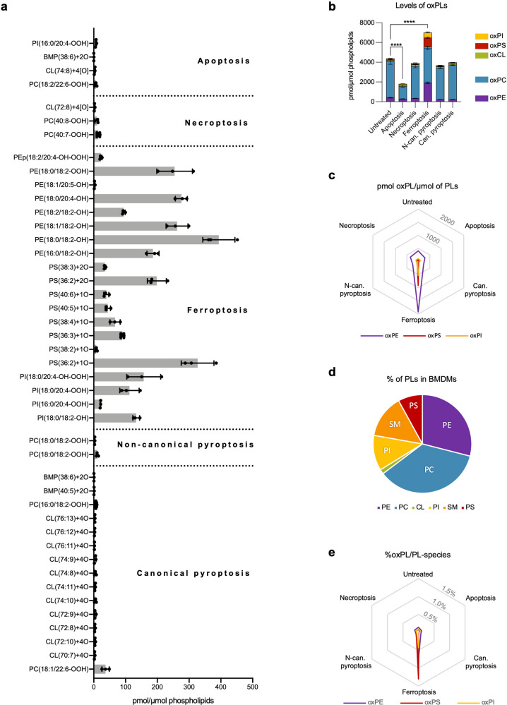
Fig. 1. Oxidative lipidomics analysis of BMDMs during apoptosis, necroptosis, ferroptosis and pyroptosis.
a “Oxidative lipidomics” of BMDMs subjected to apoptosis (4 h, 4000IU/ml hTNF and 5 μM TPCA-1), necroptosis (6 h, 4000IU/ml hTNF, 5 μM TPCA-1 and 10 μM zVAD.fmk), ferroptosis (4 h, 0.5 μM ML162), canonical pyroptosis (3 h of 1 µg/ml LPS followed by 30 min incubation with 5 mM ATP) and non-canonical pyroptosis (3 h of 1 µg/ml LPS followed by 4 h incubation after LPS transfection). Only oxidized lipid species statistically different from untreated samples are plotted. b Level of oxPLs analyzed by ‘oxidative lipidomics’ in BMDMs subjected to different types of RCD. c Level of oxPE, oxPS and oxPI in different modes of cell death. d Percentage of different groups of PL in untreated BMDMs. e Percentage of oxPLs relative to their unoxidized counterparts. The combined results of three biological replicates are shown. a, b Bars are mean ± SEM, one-way ANOVA, **p < 0.01, ****p < 0.0001.

