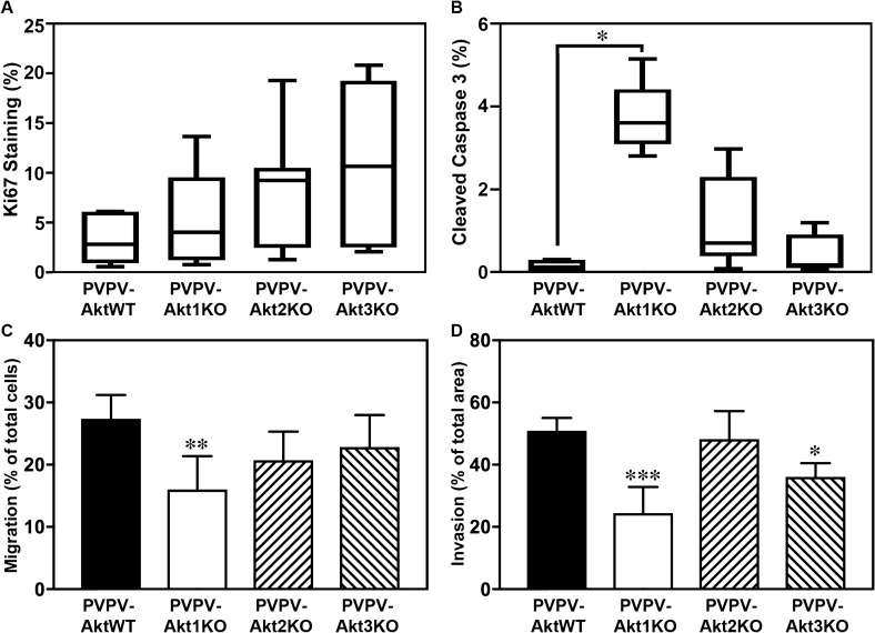
Figure 3.
Akt1 loss primarily increase thyroid cell apoptosis and reduces cell motility. Thyroid cell proliferation and apoptosis in vivo were examined by IHC using antibodies against Ki67 (A; n = 7 for all genotypes) and cleaved caspase-3 (B; n = 5 for all genotypes), respectively. Ki67 was not changed for the any of the KO mice. An increase in apoptosis was identified in the PVPV-Akt1KO thyroid glands vs PVPV-AktWT (*p = 0.008). There was a trend for an increase in PVPV-AKT2KO thyroid glands (p = 0.06). Cell migration (C) and invasion (D) of primary cultured thyroid cells in vitro were examined using Boyden chambers without or with Matrigel. PVPV-Akt1KO cells had reduced migration and PVPV-Akt1 and Akt3KO cells had reduced invasion. Cells from at least five different thyroids of each genotype mouse were tested. *p < 0.01, **p < 0.005. Graph images were created using Graphpad Prism version 8.4.2 (https://graphad.com). IHC quantitation was performed using InForm software version 2.3.0, (https://www.perkinelmer.com/Content/LST_Software_Downloads/inFormUserManual_2_3_0_rev1.pdf).

