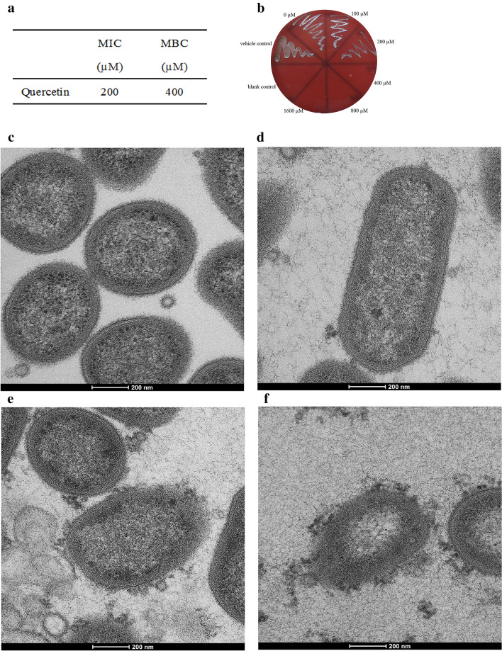
Figure 1.
Antibacterial activity and morphological changes of P. gingivalis. (a) MIC and MBC values of quercetin against planktonic P. gingivalis. 107 CFU/mL P. gingivalis suspension was added in a flat-bottomed 96-well microplate at final concentrations of 0–1600 μM quercetin and incubated under anaerobic conditions at 37 °C. (b) The number of colonies on blood agar. An aliquot of 10 μL cell suspension from each well was taken from above 96-well microplate, and bacterial clones were counted on the blood agar plates after incubation for 3–5 days. (c–f) TEM images of P. gingivalis treated with different concentration of quercetin. P. gingivalis cells with different concentrations of quercetin cultured at 37 °C for 4 h. The cell pellets were fixed with 2.5% glutaraldehyde at 4 °C, exposed to 2% osmium tetraoxide for 2 h, dehydrated in a series of ethanol and dried in acetone. Then the samples were embedded in resin blocks, cut into ultrathin sections, and stained with uranyl acetate and lead citrate. (c) 0 μM, (d) 100 μM, (e) 200 μM, (f) 400 μM, Bar = 200 nm.

