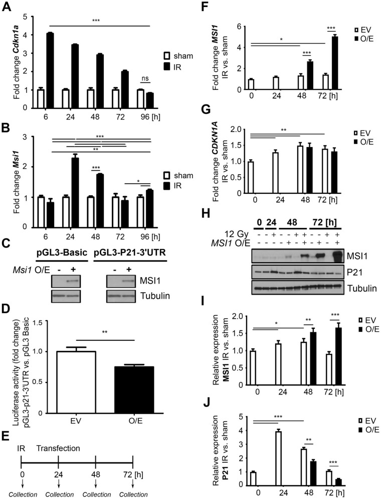
Figure 3.
MSI1 is a negative regulator of p21Waf1/Cip1 (Cdkn1a) mRNA translation. (A, B) qRT-PCR analysis of Cdkn1a (A) and Msi1 (B) in the FACS-sorted YFP+ cells isolated from the Bmi1Ctrl mice treated according to protocol 1 (Supplementary Fig. 1A). The sham mice were used as a control. IR, irradiated group (12 Gy TBI). Data are represented as the mean ± SD, n = 3 mice per group. *p < 0.05, **p < 0.01 and p < 0.001 by one-way ANOVA. (C, D) Luciferase assays of HEK293T cells transfected with the pGL3 Basic or pGL3-p21Waf1/Cip1 3′-UTR vector and Msi1 overexpression vector (pCMV6-AC-GFP-Msi1). O/E, overexpression. EV, empty vector. (C) Western blot analysis of Msi1 overexpression. Full-length blots are presented in the “Supplementary file”. (D) Relative luciferase activity. Data are represented as the mean ± SD, n = 3. **p < 0.01 by Student’s t-test. (E–J) Analysis of the effect of MSI1 on p21Waf1/Cip1 expression after γ irradiation-induced injury in vitro. HEK293T cells were seeded on a plate and irradiated with a total dose of 0 (sham) or 12 Gy (IR, irradiated), and 24 h later, MSI1 was overexpressed. Cells were collected at 0, 24, 48 and 72 h. O/E, overexpression. EV, empty vector. (E) Experimental outline. (F, G) qRT-PCR analysis of MSI1 (F) and CDKN1A (G) in HEK293T cells. (H) Western blot analysis of MSI1 and p21Waf1/Cip1 (P21) protein expression in HEK293T cells. Full-length blots are presented in the “Supplementary file”. (I, J) Densitometric analysis of protein expression in HEK293T cells performed using ImageJ software. Data are represented as the mean ± SD, n = 3. *p < 0.05, **p < 0.01 and ***p < 0.001 by Student’s t-test.

