Fig. 4.
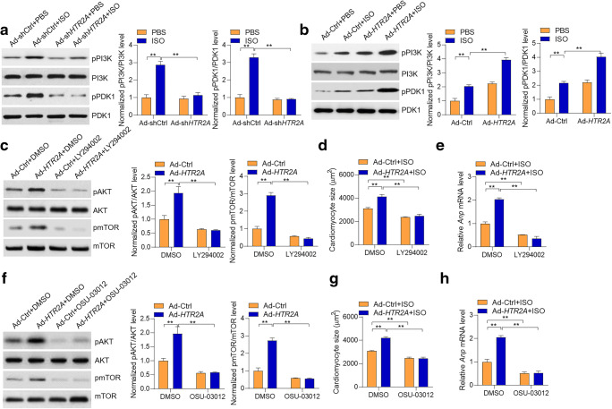
PI3K-PDK1-mediated HTR2A activation on the AKT-mTOR pathway. a HTR2A knockdown represses PI3K-PDK1 signaling. Cardiomyocytes with/without HTR2A knockdown were subjected to ISO treatment (50 μM) for 24 h. Representative western blot and quantitative results are shown (n = 3). b HTR2A overexpression promotes PI3K-PDK1 signaling. Cardiomyocytes with/without HTR2A overexpression were subjected to ISO treatment (50 μM) for 24 h. Representative western blot and quantitative results are shown (n = 3). c PI3K inhibition blocks HTR2A function in regulating AKT-mTOR signaling in cardiomyocytes. Rat cardiomyocytes with/without HTR2A overexpression were subjected to hypertrophy induction with ISO (50 μM) treatment for 24 h. The cardiomyocytes were also treated with/without PI3K inhibitor LY294002 (500 nM). d, e PI3K inhibition blocks HTR2A function in cardiomyocyte size. Rat cardiomyocytes with/without HTR2A overexpression were subjected to hypertrophy induction with ISO (50 μM) treatment for 48 h. The cardiomyocytes were also treated with/without PI3K inhibitor LY294002 (500 nM). Cardiomyocyte size was quantified with the ImageJ software (d), and the expression of hypertrophy-associated fetal genes was analyzed with qRT-PCR (e, n = 3 in each group). f PDK1 inhibition blocks HTR2A function in AKT-mTOR signaling. Rat cardiomyocytes with/without HTR2A overexpression were subjected to hypertrophy induction with ISO (50 μM) treatment for 24 h. The cardiomyocytes were also treated with/without PDK1 inhibitor OSU-03012 (2 μM). Representative western blot and quantitative results are shown (n = 3). g, h Rat cardiomyocytes with/without HTR2A overexpression were subjected to hypertrophy induction with ISO (50 μM) treatment for 48 h. The cardiomyocytes were also treated with/without PDK1 inhibitor OSU-03012 (2 μM). Cardiomyocyte size was quantified with the ImageJ software (h), and the expression of hypertrophy-associated fetal genes was analyzed with qRT-PCR (h, n = 3 in each group). **P < 0.01 analyzed by two-way ANOVA followed by the Tukey post hoc test. Cardiomyocyte culture sets were performed on three different dates. Western blot was performed for each experiment set, and representative western blot results were shown

