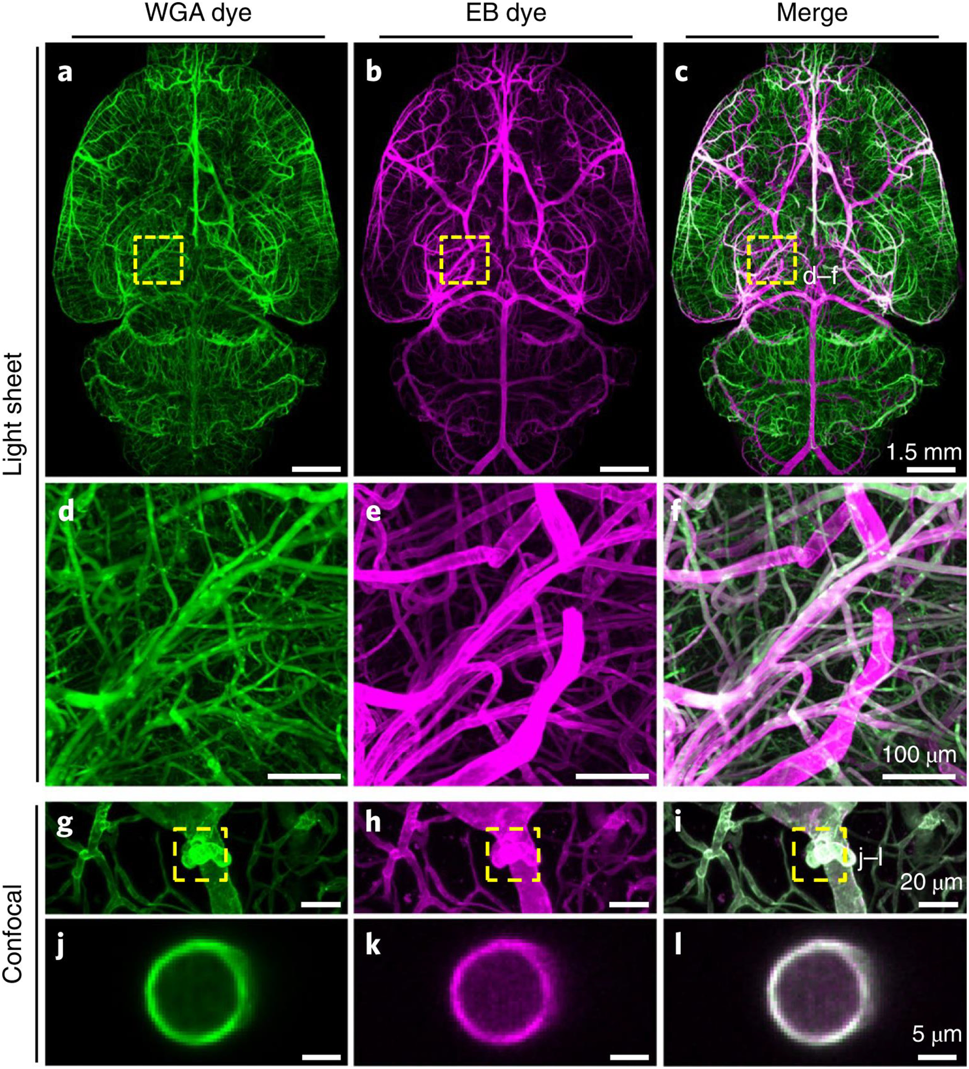
Figure 2 |. Enhancement of vascular staining using two complementary dyes.

a-c, Maximum intensity projections of the automatically reconstructed tiling scans of WGA (a) and Evans blue (b) signals in the same sample and the merged view (c). d-f: Close-up of marked region in (c). g–l, Confocal images of WGA- and EB-stained vessels and vascular wall (g–i, maximum intensity projections of 112 μm and j–l, single slice of 1 μm). The experiment was performed on 9 different mice with similar results.
