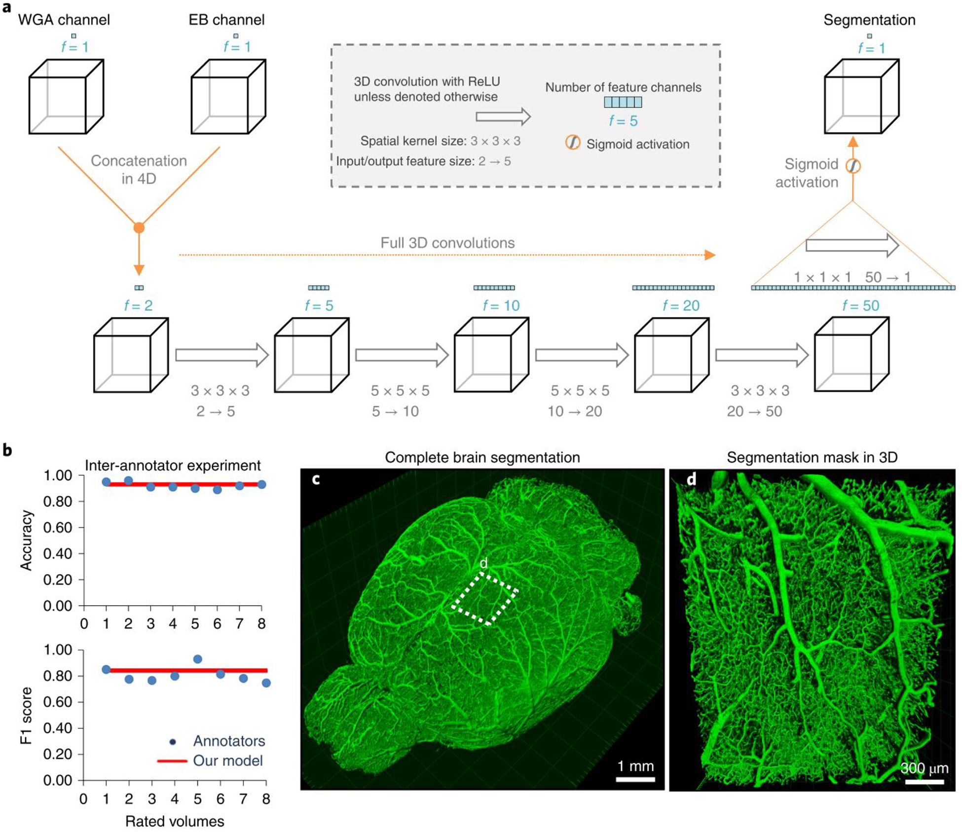
Figure 3 |. Deep learning architecture of VesSAP and performance on vessel segmentation.

a, The 3D VesSAP network architecture consisting of five convolutional layers and a sigmoid activation for the last layer including the kernel sizes and input/output of the feature size. b, The F1-score for inter-annotator experiments (blue) compared to VeSAP (red). c, 3D rendering of full brain segmentation from a CD1-E mouse. d, 3D rendering of a small volume marked in (c). The experiment was performed on 9 different mice with similar results.
