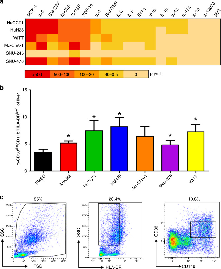
Fig. 1. Secretion of soluble factors and in vitro differentiation of immunosuppressive MDSCs by BTC cell lines.
a Heat map to summarise secretion of cytokines and chemokines from a panel of BTC cell lines. BTC cell lines were grown to 70–80% confluency, at which point supernatants were harvested and analysed using a multiplex platform. Data in the heat map represent the mean pg/mL values from at least n = 2 individual experiments. b PBMCs from healthy adult donors were incubated with 10 ng/mL of IL-6/GM-CSF (positive control) or with 10% of culture media supplemented with supernatants from individual human BTC cell lines for 7 days. These cells were then harvested, stained for MDSC phenotypic markers, and analysed via flow cytometry. Error bars represent standard deviation across donors. Asterisk (*) denotes statistical significance as compared to paired DMSO-treated PBMCs. For each condition, a minimum of n = 8 donors were analysed; Supplementary Table 3 indicates an exact n for each condition. c The gating schema for flow cytometric analysis of cells with an MDSC phenotype. Gates and voltage were set using appropriate fluorochrome-conjugated isotype control antibodies.

