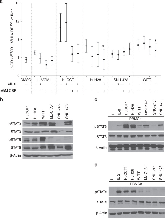
Fig. 2. Cytokine neutralization in vitro alters MDSC expansion and inhibits Jak/STAT signaling in PBMCs induced by BTC supernatants.
a PBMCs were stimulated with 10 ng/mL of IL-6/GM-CSF (positive control) or with 10% of culture media supplemented with supernatants from individual BTC cell lines ± IL-6 or GM-CSF neutralising antibody for 7 days. Cells were then harvested and stained for the MDSC phenotype and analysed via flow cytometry. Error bars represent standard error of measurement. Asterisk (*) denotes significance compared to GM-CSF neutralisation alone. For each condition, a minimum of n = 3 donors were analysed; Supplementary Table 3 indicates an exact n for each condition. b Basal activation of the Jak/STAT pathway in a panel of BTC cell lines. Immunoblot analysis was conducted to assess constitutive expression of phosphorylated STAT3 (Tyr705), STAT5 (Tyr694), and STAT1 (Tyr701). Levels of total STAT proteins and β-actin were included as loading controls. Data shown are representative from at least n = 3 individual experiments. c Healthy donor PBMCs were incubated for 20 min with culture media supplemented with 10% supernatants from individual BTC cell lines. Cells were then lysed and analysed by immunoblot. PBMCs showed increased pSTAT3 (Tyr705) and d pSTAT5 (Tyr694) following a 20-min incubation with BTC supernatants.

