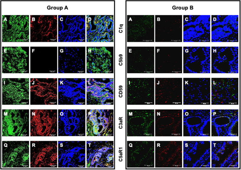
Figure 6.
C1q and C5b-9 deposition and CD59, C3a and C5a receptor expression and their co-localization with PTX3 in second biopsy of patients who developed prostate cancer (group A) or not (group B). PTX3 (A, E, I, M, Q), C1q (B), C5b-9 (F), CD59 (J), C3a (N) and C5a receptor (R) proteins were investigated by double-label immunofluorescence and confocal microscopy as detailed in Materials and Methods. TO-PRO-3 was employed to counterstain nuclei (blue; C, G, K, O, S). Merged images (yellow; D, H, L, P, T) demonstrated the co-localization (green) of C1q (D), CD59 (L), C3a (P) and C5aR1 (T) with PTX3, whereas no co-localization was evident for C5b-9 (H). Bar length = 50 µ.

