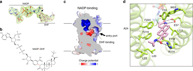
Fig. 2. Formation and binding environment of NADP–DHF adduct.
a Fo–Fc electron omit map of the NADP–DHF adduct contoured at 3σ. b Chemical structure of the NADP–DHF adduct. The covalent bond connecting the nicotinamide C-4 atom of NADPH and the C-2 atom of finasteride is highlighted in red. c Enclosed binding cavity for NADP–DHF with charge potentials. The potential entry port for the steroid substrates is indicated by an arrow. Membrane boundaries are shown as gray lines. d Molecular details of the DHF-binding pocket. Hydrogen bonds (2.2–3.2 Å) are indicated by dashed lines.

