Figure 7.
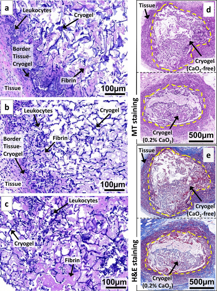
Antimicrobial microcomposite cryogels are biodegradable and elicit minimal host inflammatory responses. H&E staining of HAGM cryogel scaffold sections explanted 4 days following subcutaneous injections in the dorsal flanks of C57BL/6 mice: (a) CP-free cryogel (0% CaO2), (b) CP-containing (0.1% CaO2) cryogel, and (c) CP-containing (0.1% CaO2) cryogel contaminated with P. aeruginosa. H&E staining highlights the macroporous polymeric network of cryogels (interconnected dark blue fibers), infiltrated leukocytes (dark blue dots), fibrin formation (purple), and surrounding tissues (cryogel-free). Masson's trichrome (MT) (d) and H&E (e) staining of square-shaped implants explanted 2 months following subcutaneous injections in the dorsal flanks of C57BL/6 mice: CP-free (0% CaO2) and CP-containing (0.2% CaO2) cryogels (dimensions: 4 mm × 4 mm × 1 mm). The yellow doted lines indicate the boundary between the cryogel and the host tissue. Images are representative of n = 5 samples per condition.

