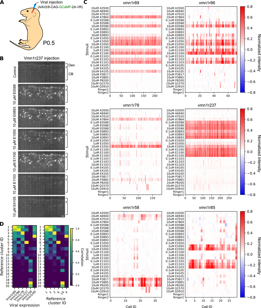
Fig. 3. Ectopic expression enabled functional analysis of vomeronasal receptors.
(A) Expression via AAV injection in the temporal vein of newborn pups. (B) Optical section from light sheet calcium imaging after the ectopic expression of GCaMP-2A-Vmn1r237 in response to different ligands. The apical layer is occupied by dendritic tips (Den) with cell bodies (CB) below. Scale bar: 20 μm (C) Calcium response after ectopic expression of GCaMP-2A-Vmn1r89, -Vmn1r86, -Vmn1r78, -Vmn1r237, -Vmn1r58, or -Vmn1r85. Ligands are identical to those in Figure 2B. (D) Pairwise correlation between the reference clustering and the ectopic responses shown in (C) (left) or autocorrelogram of the reference clustering (right).

