
Figure 6.
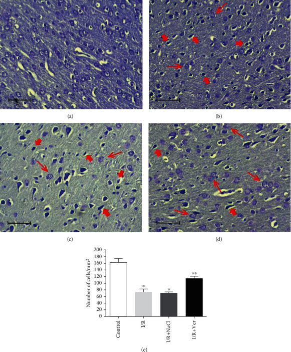
Nissl staining of rats' prefrontal cortex following transient global I/R and cell counting. The prefrontal cortex of control animals (a) did not contain any damaged neurons. However, the cortex of rats in the I/R (b) and I/R+NaCl (c) groups was characterized by fewer Nissl stain neurons (thin arrows) than that in the I/R+Ver group (d) and markedly contained shrunken, intensely stained, and dystrophic neurons (thick arrows). As shown in the graph (e), treatment rats with verapamil significantly reduced neuronal cell loss in the prefrontal cortex. Data are represented as mean ± SD. Scale bars 100 μm, magnification ×400. ∗P < 0.001 vs. control group, ∗∗P < 0.001 vs. I/R+NaCl and I/R groups.