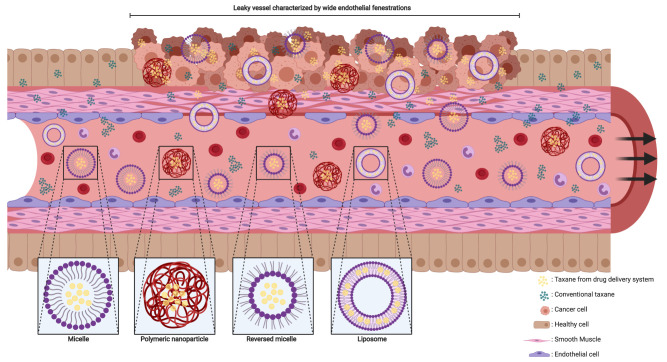
Figure 2.
Schematic impression of EPR effect and nanoparticles. The fenestrae in the vascular wall of the tumor are wider than in normal tissue and the smooth muscle cells in the vascular wall are arranged in a chaotic manner compared to healthy tissue. Nanoparticles and conventional taxanes can easily pass the vascular wall inside the tumor. In contrast, the endothelial cells and smooth muscle cells are good aligned in the vascular wall of healthy tissue, which makes it hard for nanoparticle to cross the wall while conventional taxanes can still penetrate inside healthy tissue. This figure was created with BioRender.com.

