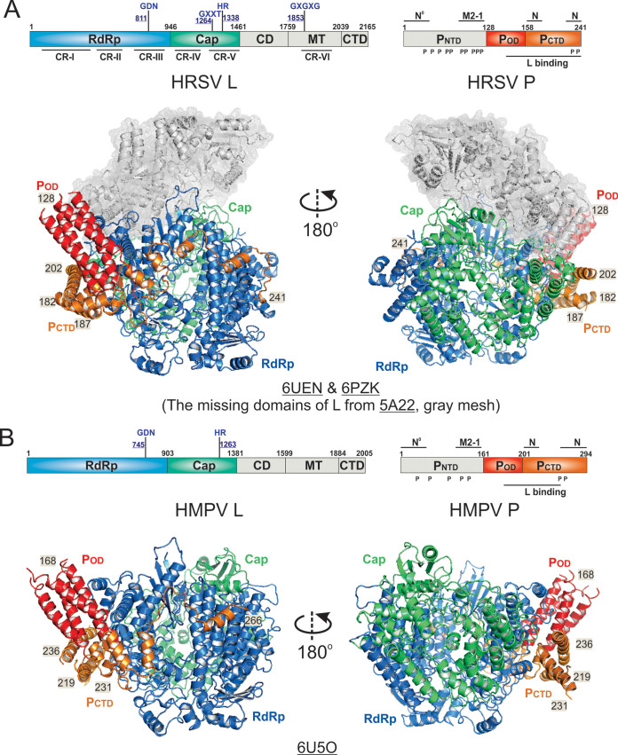
FIG 4.
The cryo-EM structures of the Pneumoviridae polymerases. (A) Linear domain representation of the L and P proteins of the human respiratory syncytial virus (HRSV) polymerase. The cartoon view of the 3.67-Å (PDB: 6UEN) and 3.2-Å (PDB: 6PZK) cryo-EM structures of HRSV polymerase complexes. The missing domains compared with the VSV L are shown in the gray meshes. (B) Linear domain representation of the L and P proteins of the human metapneumovirus (HMPV) polymerase. The cartoon view of the 3.7-Å (PDB: 6U5O) cryo-EM structure of the HMPV polymerase is shown. The domain colorings are the same as Fig. 2. The terminal residue numbers of the modeled POD and PCTD are indicated. The PDB accession codes are underlined.

