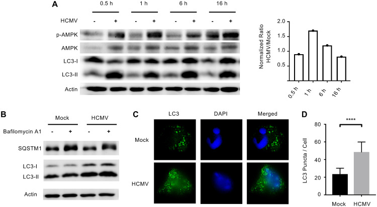
FIG 1.
HCMV infection induces autophagy in monocytes. (A) Human peripheral blood monocytes were mock or HCMV infected for 30 min or 1, 6, or 16 h. Levels of phosphorylated AMPK (p-AMPK), total AMPK, LC3-I, LC3-II, or actin were detected by immunoblotting from whole-cell lysates. Levels of p-AMPK were normalized to actin and then total AMPK for each treatment and time point. The ratio of HCMV to mock of these values was then plotted for each time point (right). (B) Mock- or HCMV-infected monocytes were treated with 200 nM bafilomycin A1 for 6 h and the levels of SQSTM1 and LC3 were determined by immunoblot. (C and D) Monocytes were mock or HCMV infected for 6 h, followed by immunofluorescent analysis with anti-LC3 antibody (green) or DAPI (blue). Results are representative of at least 3 independent experiments using monocytes from different blood donors. (D) LC3 puncta per cell were counted using FIJI and the average puncta per cell were plotted with the mean and the 95% confidence interval (CI); ****, P < 0.0005.

