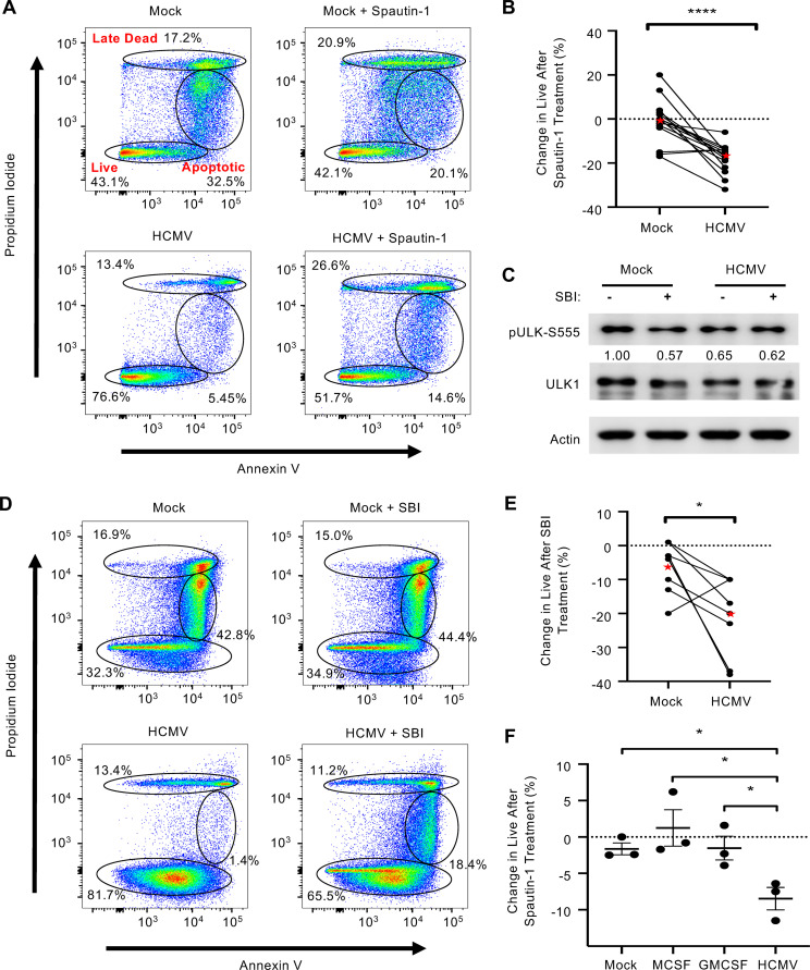
FIG 3.
HCMV-induced autophagy stimulates survival of infected monocytes. (A and D) Human peripheral blood monocytes were mock or HCMV infected for 24 h, followed by treatment with either 50 μM Spautin-1, 10 μM SBI-0206965 (SBI), or vehicle control for an additional 24 h. Viability was measured by flow cytometry using propidium iodide (PI) and annexin V staining. Gates represent live, apoptotic, and late dead cells. (B, E, and F) Change in survival after the addition of Spautin-1 or SBI was quantified and plotted for mock- and HCMV-infected monocytes (B and E) or myeloid growth factors MCSF and GMCSF (F). Lines connect donor-matched data points from the same experiment. Red stars in B and E indicate the mean result for each group. Results in E are plotted as the mean ± SEM. (C) Levels of pULK1-S555 and ULK1 were determined by immunoblotting in the presence or absence of SBI in HCMV- or mock-infected monocytes after 6 h. All results are representative of at least 3 independent experiments using different donors; ****, P < 0.0005; *, P < 0.05.

