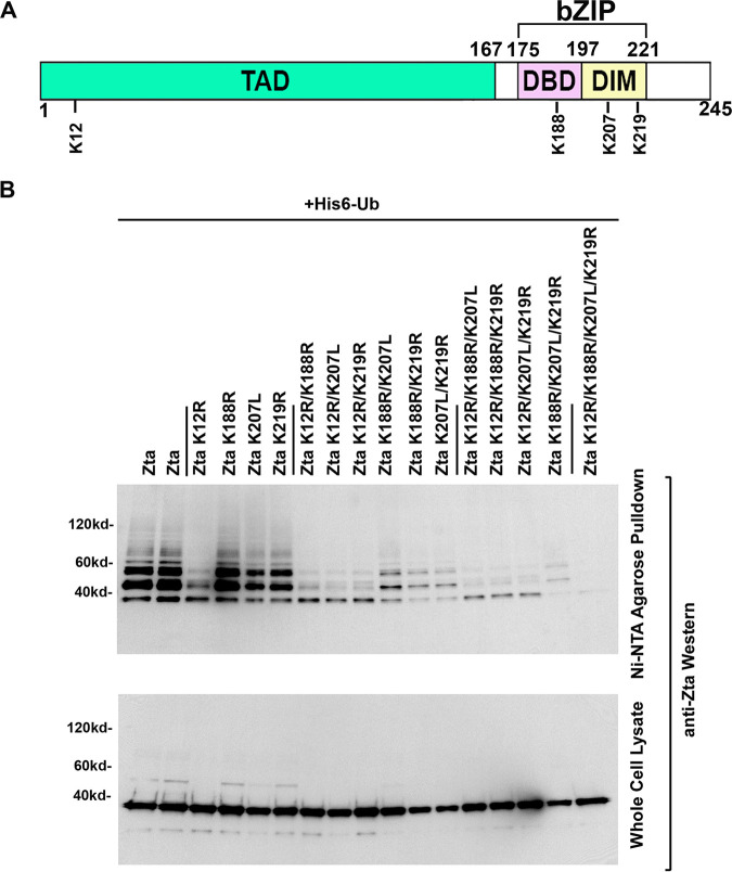
FIG 8.
Analysis of multiple-lysine-residue Zta mutants. (A) Schematic diagram of selected Zta lysine residues. (B) EBV-negative AdAH cells were transfected with 5 μg of a ubiquitin (His6-Ub) expression vector and 5 μg of the indicated Zta or Zta mutant expression plasmids. Cells were lysed under denaturing conditions and extracts were purified over nickel-NTA beads. The purified material was assayed by Western blot analysis employing anti-Zta antibodies. Expression levels of Zta and its mutants were determined by Western blot analysis of whole-cell lysates.

