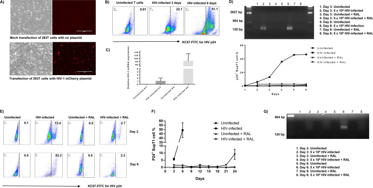
FIG 1.
HIV infection of SupT1 cells. (A) Bright and fluorescence microscopic examinations of HEK293T cells with mock transfection (top) and transfected for 48 h with HIV NL4-3 mCherry plasmid (bottom). (B) Flow cytometry analysis of p24 expression in SupT1 cells with HIV infection. (C) Reverse transcription-PCR (RT-PCR) analysis of HIV-1 mRNA levels in SupT1 cells with or without HIV infection on day 3 and day 6. (D) RT-PCR detection of HIV gene expression in SupT1 cells with HIV infection and raltegravir (RAL) treatment (added 2 h post HIV infection). (E) Flow cytometry analysis of p24 expression in SupT1 cells with HIV infection and RAL treatment (added 24 h post HIV infection). (F) Time-dependent p24 expression in SupT1 cells with HIV infection and RAL treatment (added 24 h post HIV infection). (G) RT-PCR detection of HIV gene expression in SupT1 cells with HIV infection and RAL treatment (added 24 h post HIV infection).

