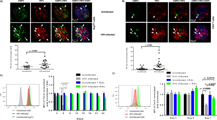
FIG 5.
DNA damage response in HIV-infected SupT1 and CD4 T cells with or without RAL treatment. (A and B) Confocal microscopy analysis of 53BP1 and TRF1 colocalization as a marker for dysfunctional telomere-induced foci (TIF) in SupT1 and primary CD4 T cells with HIV infection. The confocal images were analyzed using the confocal software Leica Application Suite X (LAS X), and summary data for the colocalization of TRF1 and 53B1 signal analyzed in each microscopic frame are shown. (C and D) Representative overlaid histograms and summary data of mean fluorescence intensity (MFI) of Tel-C in SupT1 cells or primary CD4 T cells, with or without HIV infection and RAL treatment, as measured by flow-FISH at the indicated times.

