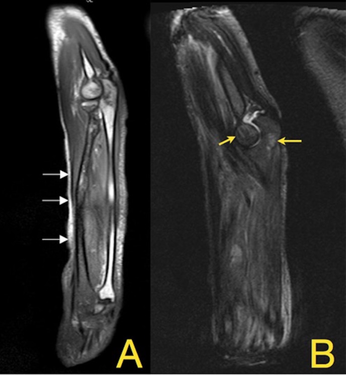
Figure 4.
(A) Coronal T1 MRI of the right forearm shows abnormal marrow signals (arrows) with restricted diffusion on the right radius extending from its proximal head to the distal radial epiphysial aspects associated with cortical erosive and lytic changes. This is in contrast to the normal ulnar bone on the right side of the image. (B) T2 MRI shows hyperintense lesions (yellow arrows) and abnormal marrow signals on the olecranon process and the humeral epicondyle.

