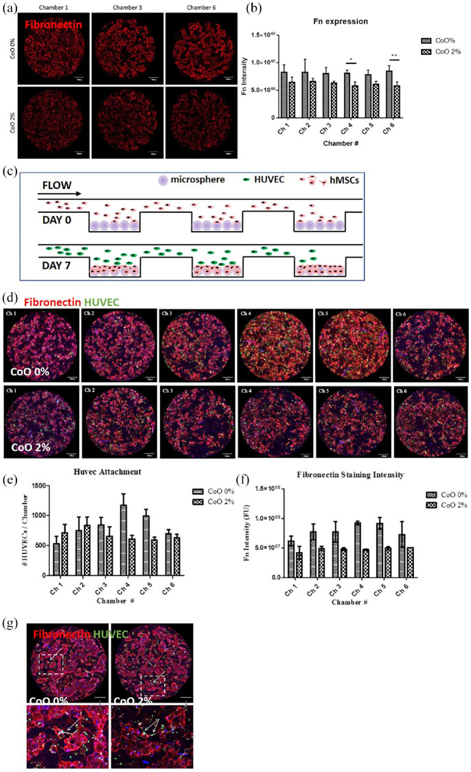
Figure 8.
Characterisation of hMSC-endothelial cell interactions using the microfluidic device. (a) Extracellular fibronectin deposition by hMSCs was assessed after 7 days under perfusion and (b) a tendency of greater deposition in response to CoO 0% was observed. (c) Representative images showing HUVEC attachment to hMSC-microsphere material. Quantification indicated that (d) HUVEC attachment to 7 day hMSC-microsphere cultures after 24 h was increased on CoO 0% PG microspheres compared to CoO 2% PG microspheres in downstream chambers (4–5), associated with corresponding fibronectin deposition (e). (f) Extended culture of HUVECs on the hMSC-microspheres for a further 48 h (to a total of 3 days culture) revealed interactions between HUVECs and the hMSC-microsphere clusters including limited formation of tubule-like structures (indicated by arrows). For (b) Mean ± SD, n = 4; For (d and e), pooled data from two independent experiments are shown as Mean ± SD, n = 2. Scale bars = 500 µm.

