
Fig 3.
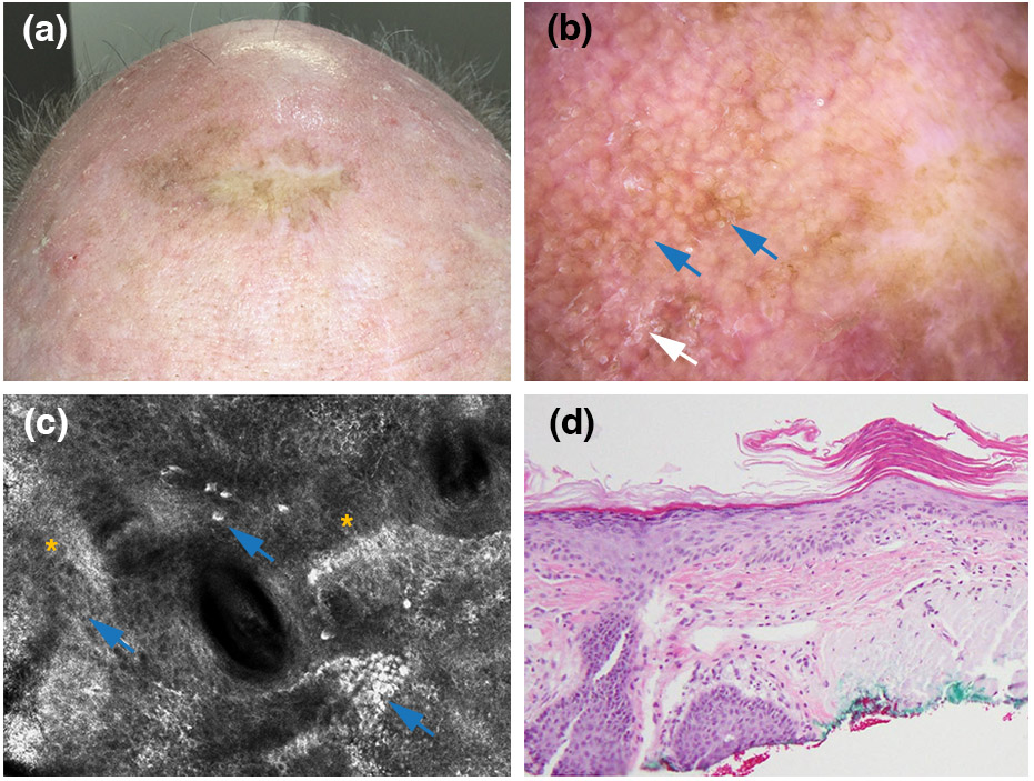
Pigmented actinic keratosis arising within a lentigo maligna surgical scar. (a) Clinical picture showing pigment extends beyond the graft margins. (b) Dermoscopic findings, showing a pseudonetwork (blue arrows) and scale (white arrow) (polarized light dermoscopy; original magnification, 10X). (c) Reflectance confocal microscopy findings, showing pigmented keratinocytes (blue arrows) and an overall atypical honeycomb pattern (yellow asterisks) (750 x 750 μm). (d) Histopathologically confirmed pigmented actinic keratosis (haematoxylin and eosin; magnification, 20X).