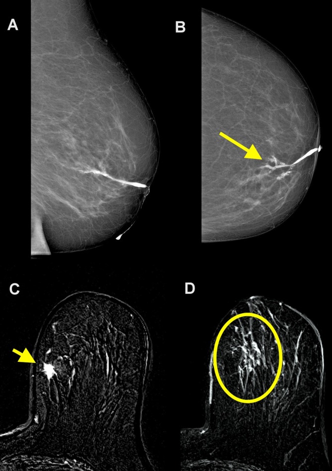
Figure 3.

A 75-year-old female with 6 months of pathological discharge from the left nipple. Duct ectasia was diagnosed on ultrasound. No abnormal findings were found on mammography. Cytology of discharge showed amorphous material, red cells, and macrofages without ductal cell. Galactography allowed for a good visualization of the main duct with a filling stop for secondary ducts (arrow) (A, B). Contrast-enhanced subtracted axial MR images show an enhancing spiculated mass at the upper inner quadrant of left breast (arrow in C) associated with a segmental non-mass enhancement (circle in D). At pathology, an invasive ductal carcinoma with extensive intraductal component was revealed.
