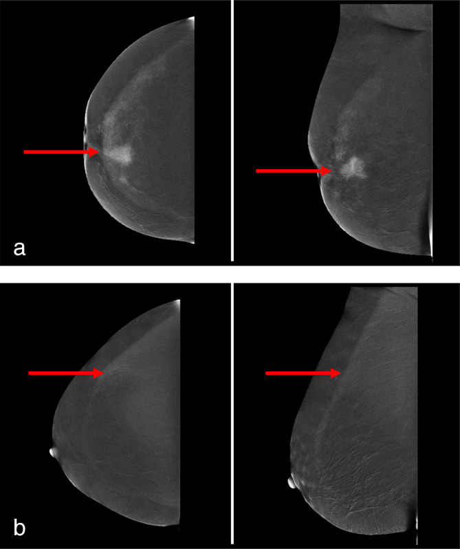
Figure 1.

(a) CEM images of a female patient with a 25 mm large invasive lobular carcinoma in her left breast, respectively on CC (left image) and MLO (right image). The red arrows demonstrate the suspicious lesion, which was considered strong enhancement by all three readers. (b) CEM images of a female patient with a 10 mm large invasive lobular carcinoma in her left breast, respectively on CC (left image) and MLO (right image). The red arrows demonstrate the suspicious lesion, which was considered weak enhancement by all three readers.
