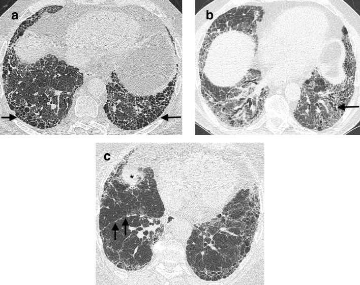
Figure 1.
CT signs of lung fibrosis. Honeycomb cysts (arrow) lying in layers in the peripheral basal aspect of the lower lobes (a). Dilated varicose tortuous airways representing traction bronchiectasis (arrow) lying amidst dense fibrotic lung in the lower lobes (b). Volume loss in the lower lobes in a patient with idiopathic pulmonary fibrosis (c). In health, on axial CT images the most inferior aspect of the oblique fissures should reach the anterior chest wall at the level of the hemidiaphragms. However, in this patient, the right oblique fissure (arrows) has been pulled back and now only reaches the midpoint of the lung at a level where the right hemidiaphragm is visible (asterix).

