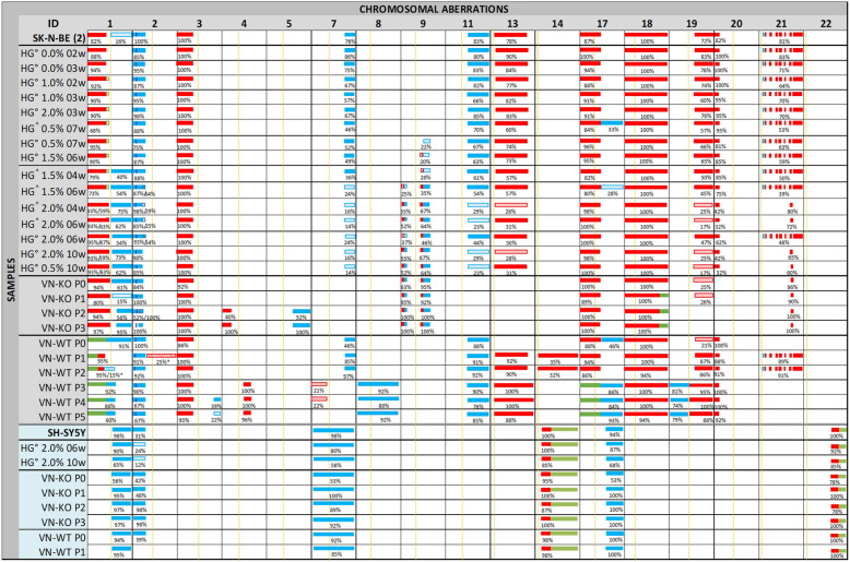
Fig. 1.
Schematic representation of the chromosomal aberrations detected by HD-SNPa in samples derived from SK-N-BE(2) and SH-SY5Y cell lines. SK-N-BE(2) and SH-SY5Y ID (identified by grey and blue columns, respectively) refer to the cell line cultured in 2D in vitro acquired from the ATCC. The presence of aberrations in chromosome 9 of the SK-N-BE(2) cell line grown in hydrogels alone and in co-culture with Schwann cells marks its organization within the figure. HG° and HG^= neuroblasts cultured in hydrogels only and coculture with Schwann cells, respectively, followed by the percentage of AlgMA (%) and the weeks of culture (w). Tumor samples from the experimental RAG1−/− VN−/− mice (VN-KO) and control RAG1−/−VN+/+ mice (VN-WT) are followed by the passage number (P0-P5). The lack of symmetry of the SH-SY5Y and SK-N-BE(2) groups is due to the absence of new changes in chromosomal aberrations in samples derived from the SH-SY5Y cell line, and to the P1 growth stop of VN-WT tumors derived from this cell line. For each altered chromosome, the approximate position of centromere is marked with a yellow line. Heterozygotic gains of genomic material (3 copies) are represented in blue, heterozygotic deletions of genomic material (1 copy) are in red, and CNLOH in green. MYCN amplification is represented with a purple line. The percentages of cells having each chromosomal aberration according to their smooth signal are below the representative bars (e.g. when we estimated a median copy number state across a segmental chromosome aberration -SCA- of 2.62 using ChAS, we inferred the SCA affected 62% of cultured cells, and median copy number state of 1.62, implying that the deletion affected 38% of the cells in the sample [41]). When the percentage is less than 30% the background color of the aberration is lighter. The striped background and the asterisk (*) after cell percentage indicates aberrations only found in the HD-SNPa of ctDNA

